CD163抗体
产品名称: CD163抗体
英文名称: CD163
产品编号: 2527
产品价格: null
产品产地: 上海
品牌商标: 雅吉
更新时间: null
使用范围: WB ELISA IHC-P IHC-F Flow-Cyt ICC IF
上海雅吉生物科技有限公司
- 联系人 :
- 地址 : 上海市闵行区元江路5500号第1幢5658室
- 邮编 :
- 所在区域 : 上海
- 电话 : 158****3937 点击查看
- 传真 : 点击查看
- 邮箱 : yajikit@163.com
| 中文名称 | CD163抗体 |
| 别 名 | Scavenger receptor cysteine-rich type 1 protein M130; CD 163; CD163 antigen; CD163 molecule; Hemoglobin Scavenger Receptor; M130; M130 antigen precursor; Macrophage associated antigen; MM130; C163A_HUMAN. |
| 研究领域 | 心血管 免疫学 细胞膜受体 |
| 抗体来源 | Rabbit |
| 克隆类型 | Polyclonal |
| 交叉反应 | Human, Mouse, (predicted: Rat, Dog, Pig, Horse, ) |
| 产品应用 | WB=1:500-2000 ELISA=1:500-1000 IHC-P=1:100-500 IHC-F=1:100-500 Flow-Cyt=3ug/Test ICC=1:100-500 IF=1:100-500 (石蜡切片需做抗原修复) not yet tested in other applications. optimal dilutions/concentrations should be determined by the end user. |
| 分 子 量 | 130kDa |
| 细胞定位 | 细胞膜 分泌型蛋白 |
| 性 状 | Liquid |
| 浓 度 | 1mg/ml |
| 免 疫 原 | KLH conjugated synthetic peptide derived from human CD163:1001-1121/1156 |
| 亚 型 | IgG |
| 纯化方法 | affinity purified by Protein A |
| 储 存 液 | 0.01M TBS(pH7.4) with 1% BSA, 0.03% Proclin300 and 50% Glycerol. |
| 保存条件 | Shipped at 4℃. Store at -20 °C for one year. Avoid repeated freeze/thaw cycles. |
| PubMed | PubMed |
| 产品介绍 | CD163 is a 130 kDa membrane glycoprotein. It is a member of the scavenger receptor cysteine-rich superfamily and is a receptor for the hemoglobin-haptoglobin complex. CD163 is expressed exclusively on the cell surface of human monocytes and macrophages that evolve predominantly in the late phase of inflammation. CD163 is present on all CD14 positive monocytes, most CD64 positive monocytes, and shows higher expression on CD16 positive monocytes. CD163 is upregulated on mononuclear phagocytes by IL-10, IL-6 and dexamethasone. Lipopolysaccharide (LPS) and phorbol myristate acetate (PMA) both induce shedding of CD163 from the cell surface into plasma or cell supernatant. Function: Acute phase-regulated receptor involved in clearance and endocytosis of hemoglobin/haptoglobin complexes by macrophages and may thereby protect tissues from free hemoglobin-mediated oxidative damage. May play a role in the uptake and recycling of iron, via endocytosis of hemoglobin/haptoglobin and subsequent breakdown of heme. Binds hemoglobin/haptoglobin complexes in a calcium-dependent and pH-dependent manner. Exhibits a higher affinity for complexes of hemoglobin and multimeric haptoglobin of HP*1F phenotype than for complexes of hemoglobin and dimeric haptoglobin of HP*1S phenotype. Induces a cascade of intracellular signals that involves tyrosine kinase-dependent calcium mobilization, inositol triphosphate production and secretion of IL6 and CSF1. Isoform 3 exhibits the higher capacity for ligand endocytosis and the more pronounced surface expression when expressed in cells. Subcellular Location: Secreted and Cell membrane. Isoform 1 and isoform 2 show a lower surface expression when expressed in cells. Tissue Specificity: Expressed in monocytes and mature macrophages such as Kupffer cells in the liver, red pulp macrophages in the spleen, cortical macrophages in the thymus, resident bone marrow macrophages and meningeal macrophages of the central nervous system. Expressed also in blood. Isoform 1 is the lowest abundant in the blood. Isoform 2 is the lowest abundant in the liver and the spleen. Isoform 3 is the predominant isoform detected in the blood. Post-translational modifications: A soluble form (sCD163) is produced by proteolytic shedding which can be induced by lipopolysaccharide, phorbol ester and Fc region of immunoglobulin gamma. This cleavage is dependent on protein kinase C and tyrosine kinases and can be blocked by protease inhibitors. The shedding is inhibited by the tissue inhibitor of metalloproteinase TIMP3, and thus probably induced by membrane-bound metalloproteinases ADAMs. Phosphorylated. Similarity: Contains 9 SRCR domains. SWISS: Q86VB7 Gene ID: 9332 Database links: Entrez Gene: 9332 Human Omim: 605545 Human SwissProt: Q86VB7 Human Unigene: 504641 Human Important Note: This product as supplied is intended for research use only, not for use in human, therapeutic or diagnostic applications. CD163是SRCR超家族成员之一,又称为血红蛋白清道夫受体(hemoglobin scavenger receptor, HbSR)、M130或p155。CD163的表达水平受很多因素调节,糖皮质激素及抗炎介质如IL-10以及IL-6能上调CD163, 而促炎介质如脂多糖(lipopolysaccharide, LPS)、γ-干扰素(interferon-γ, IFN-γ)以及TNF-α则抑制CD163表达。 CD163是一种I型膜蛋白,又称为M130抗原、Ber-Mac3、Ki-M8或SM4。CD163限制性表达于单核/巨噬细胞系,所有循环系统的单核细胞和大多数组织(除外淋巴滤泡套区和生发中心)的巨噬细胞均阳性表达,主要用于单核/巨噬细胞的检测。 |
| 产品图片 | 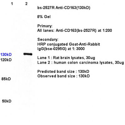 Sample: Sample:Lane1: Brain (Rat) Lysate at 30 ug Lane2: Colon carcinoma(Human) Lysate at 30 ug Primary: Anti- CD163 (bs-2527R) at 1/300 dilution Secondary: IRDye800CW Goat Anti-Rabbit IgG at 1/20000 dilution Predicted band size: 130 kD Observed band size: 130kD 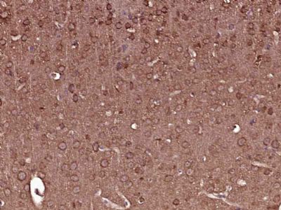 Paraformaldehyde-fixed, paraffin embedded (Mouse brain); Antigen retrieval by boiling in sodium citrate buffer (pH6.0) for 15min; Block endogenous peroxidase by 3% hydrogen peroxide for 20 minutes; Blocking buffer (normal goat serum) at 37°C for 30min; Antibody incubation with (CD163) Polyclonal Antibody, Unconjugated (bs-2527R) at 1:400 overnight at 4°C, followed by operating according to SP Kit(Rabbit) (sp-0023) instructionsand DAB staining. Paraformaldehyde-fixed, paraffin embedded (Mouse brain); Antigen retrieval by boiling in sodium citrate buffer (pH6.0) for 15min; Block endogenous peroxidase by 3% hydrogen peroxide for 20 minutes; Blocking buffer (normal goat serum) at 37°C for 30min; Antibody incubation with (CD163) Polyclonal Antibody, Unconjugated (bs-2527R) at 1:400 overnight at 4°C, followed by operating according to SP Kit(Rabbit) (sp-0023) instructionsand DAB staining.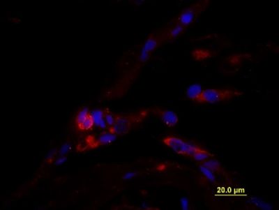 Formalin-fixed and paraffin embedded Human testis tissue labeled with unconjugated Anti-CD163/M130 Polyclonal Antibody, unconjugated (bs-2527R) at 1:100 for 40 minutes at 37°C followed by labeling Donkey Anti-Rabbit, Cy3 conjugated 1:300, 60 minutes at 37°C. DAPI nuclear stain employed. Image shows membrane staining of testicular macrophages in the interstitial compartment of the testis, while cells in the seminiferous tubules are negative. Formalin-fixed and paraffin embedded Human testis tissue labeled with unconjugated Anti-CD163/M130 Polyclonal Antibody, unconjugated (bs-2527R) at 1:100 for 40 minutes at 37°C followed by labeling Donkey Anti-Rabbit, Cy3 conjugated 1:300, 60 minutes at 37°C. DAPI nuclear stain employed. Image shows membrane staining of testicular macrophages in the interstitial compartment of the testis, while cells in the seminiferous tubules are negative. Blank control (Black line): U2OS. Blank control (Black line): U2OS.Primary Antibody (green line): Rabbit Anti-CD163 antibody (bs-2527R) Dilution: 3μg /10^6 cells; Isotype Control Antibody (orange line): Rabbit IgG . Secondary Antibody (white blue line): Goat anti-rabbit IgG-PE Dilution: 1μg /test. Protocol The cells were fixed with 4% paraformaldehyde for 10 min at room temperature. Cells incubated in 5%BSA to block non-specific protein-protein interactions for 30 min at room temperature. The cells were then stained with Primary Antibody for 30 min at room temperature. The secondary antibody used for 40 min at room temperature. Acquisition of 20,000 events was performed. |