阶段特异性胚胎表面抗原-1抗体
产品名称: 阶段特异性胚胎表面抗原-1抗体
英文名称: Wnt3a
产品编号: 1700
产品价格: null
产品产地: 上海
品牌商标: 雅吉
更新时间: null
使用范围: WB ELISA IHC-P IHC-F Flow-Cyt IF
- 联系人 :
- 地址 : 上海市闵行区元江路5500号第1幢5658室
- 邮编 :
- 所在区域 : 上海
- 电话 : 158****3937 点击查看
- 传真 : 点击查看
- 邮箱 : yajikit@163.com
| 中文名称 | 阶段特异性胚胎表面抗原-1抗体 |
| 别 名 | Fut4; SSEA-1; 3-FAL; Alpha (1,3) fucosyltransferase; Alpha 13 fucosyltransferase FucT; EC 2.4.1.; ELAM 1 ligand fucosyltransferase; ELAM ligand fucosyltransferase; ELAM1 ligand fucosyltransferase; ELFT; FCT3A; Fuc TIV; Fucosyltransferase 4 alpha 1 3 fucosyltransferase myeloid specific; Fucosyltransferase 4; FucT IV; FUCTIV; FUT4; Galactoside 3 L fucosyltransferase; Lewis X; LeX; SSEA 1; SSEA1; Stage specific embryonic antigen 1; FUC-TIV. |
研究领域肿瘤 细胞生物 免疫学 细胞膜受体
抗体来源Rabbit
克隆类型Polyclonal
交叉反应Human, Mouse, (predicted: Rat, )
产品应用WB=1:500-2000 ELISA=1:500-1000 IHC-P=1:100-500 IHC-F=1:100-500 Flow-Cyt=1μg/Test IF=1:100-500 (石蜡切片需做抗原修复)
not yet tested in other applications.
optimal dilutions/concentrations should be determined by the end user.
分 子 量58kDa
细胞定位细胞浆 细胞膜
性 状Liquid
浓 度1mg/ml
免 疫 原KLH conjugated synthetic peptide derived from human CD15:251-295/433
亚 型IgG
纯化方法affinity purified by Protein A
储 存 液0.01M TBS(pH7.4) with 1% BSA, 0.03% Proclin300 and 50% Glycerol.
保存条件Shipped at 4℃. Store at -20 °C for one year. Avoid repeated freeze/thaw cycles.
PubMedPubMed
产品介绍The Lewis histo-blood group system comprises a set of fucosylated glycosphingolipids that are synthesized by exocrine epithelial cells and circulate in body fluids. The glycosphingolipids function in embryogenesis, tissue differentiation, tumor metastasis, inflammation, and bacterial adhesion. They are secondarily absorbed to red blood cells giving rise to their Lewis phenotype. This gene is a member of the fucosyltransferase family, which catalyzes the addition of fucose to precursor polysaccharides in the last step of Lewis antigen biosynthesis. It encodes an enzyme with alpha(1,3)-fucosyltransferase and alpha(1,4)-fucosyltransferase activities. Mutations in this gene are responsible for the majority of Lewis antigen-negative phenotypes. Multiple alternatively spliced variants, encoding the same protein, have been found for this gene. [provided by RefSeq].
Function:
May catalyze alpha-1,3 glycosidic linkages involved in the expression of Lewis X/SSEA-1 and VIM-2 antigens.
Subcellular Location:
Golgi apparatus, Golgi stack membrane; Single-pass type II membrane protein. Note=Membrane-bound form in trans cisternae of Golgi.
Tissue Specificity:
Highest expression in stomach and colon. It is also expressed in the lung, testis, uterus, small intestine and to a lesser extent in spleen, and ovary. Present in trace amounts in brain, thymus, heart, smooth muscle, kidney and bone marrow. Not found in liver, salivary gland and pancreas.
Similarity:
Belongs to the glycosyltransferase 10 family.
SWISS:
Q11127
Gene ID:
2526
Database links:
Entrez Gene: 10690 Human
Entrez Gene: 2526 Human
Entrez Gene: 2527 Human
Entrez Gene: 2528 Human
Entrez Gene: 2529 Human
Omim: 104230 Human
SwissProt: P22083 Human
SwissProt: P51993 Human
SwissProt: Q11128 Human
SwissProt: Q11130 Human
SwissProt: Q9Y231 Human
Unigene: 390420 Human
Unigene: 457 Human
Unigene: 49117 Human
Unigene: 572064 Human
Unigene: 623098 Human
Unigene: 631843 Human
Unigene: 631846 Human
Unigene: 705615 Human
Important Note:
This product as supplied is intended for research use only, not for use in human, therapeutic or diagnostic applications.
CD15(SSEA-1)是胚胎干细胞表达胚胎阶段特异性抗原的主要SSEA-1蛋白,还包括、SSEA-3、TRA-1-81、TRA-1-60等。CD15主要存在于粒细胞(包括中性粒细胞和嗜酸性粒细胞)以及部分单核细胞上。它在介导细胞吞噬细菌和趋化性方面起重要作用。CD15常表达于霍奇金氏病中R-S细胞,尤其是经典型霍奇金淋巴瘤中。结节硬化型霍奇金淋巴瘤多为CD15阳性。CD15抗体是鉴定霍奇金氏病的一个有效工具。
| 产品图片 |
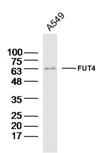 Sample:A549 Cell (Human) Lysate at 40 ug
Primary: Anti-CD15(bs-1702R)at 1/300 dilution Secondary: IRDye800CW Goat Anti-Rabbit IgG at 1/20000 dilution Predicted band size: 58kD Observed band size: 63kD 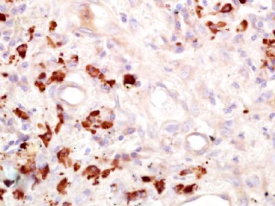 Paraformaldehyde-fixed, paraffin embedded (Human lung cancer); Antigen retrieval by boiling in sodium citrate buffer (pH6.0) for 15min; Block endogenous peroxidase by 3% hydrogen peroxide for 20 minutes; Blocking buffer (normal goat serum) at 37°C for 30min; Antibody incubation with (CD15) Polyclonal Antibody, Unconjugated (bs-1702R) at 1:400 overnight at 4°C, followed by operating according to SP Kit(Rabbit) (sp-0023) instructionsand DAB staining.
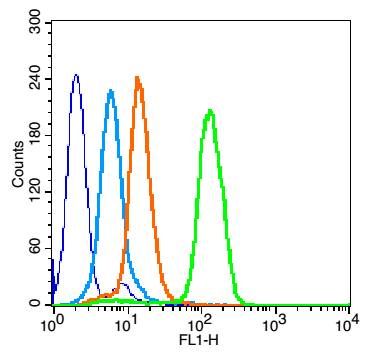 Overlay histogram showing HL 60 cells stained with bs-1702R (Green line).
The cells were fixed with 90% methanol (5 min) and then permeabilized with 0.01M PBS-Tween for 20 min. The cells were then incubated in 1x PBS / 10% normal goat serum to block non-specific protein-protein interactions followed by the antibody (bs-1702R,1μg/1x10^6 cells) for 30 min at 22℃. The secondary antibody used was fluorescein isothiocyanate goat anti-rabbit IgG (H+L) (bs- 0295G-FITC , Brillant blue line) at 1/200 dilution for 30 min at 22℃. Isotype control antibody was rabbit IgG (polyclonal,bs-0295P,Orange line) (1μg/1x10^6 cells) used under the same conditions. Unlabelled sample (blue line) was also used as a control. Acquisition of 20,000 events were collected using a 20mW Argon ion laser (488nm) and 525/30 bandpass filter. 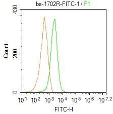 Blank control: Mouse spleen.
Primary Antibody (green line): Rabbit Anti-CD15 /FITC Conjugated antibody (bs-1702R-FITC) Dilution: 1μg /10^6 cells; Isotype Control Antibody (orange line): Rabbit IgG-FITC . Protocol The cells were fixed with 4% PFA (10min at room temperature)and then permeabilized with 0.1% PBST for 20 min at-20℃. The cells were then incubated in 5% BSA to block non-specific protein-protein interactions for 30 min at room temperature. The cells were stained with Primary Antibody for 30 min at room temperature. Acquisition of 20,000 events was performed. |