血管内皮细胞粘附分子抗体
产品名称: 血管内皮细胞粘附分子抗体
英文名称: VCAM-1
产品编号: 0396
产品价格: null
产品产地: 上海
品牌商标: 雅吉
更新时间: null
使用范围: WB ELISA IHC-P IHC-F Flow-Cyt IF
- 联系人 :
- 地址 : 上海市闵行区元江路5500号第1幢5658室
- 邮编 :
- 所在区域 : 上海
- 电话 : 158****3937 点击查看
- 传真 : 点击查看
- 邮箱 : yajikit@163.com
| 中文名称 | 血管内皮细胞粘附分子抗体 |
| 别 名 | VCAM1; CD 106; CD106; CD106 Antigen; DKFZp779G2333; INCAM 100; L1CAM; MGC99561; Vascular Cell Adhesion Molecule 1; Vascular cell adhesion protein 1; VCAM 1; VCAM-1; INCAM-100; V-CAM 1; VCAM1_HUMAN. |
研究领域 肿瘤 心血管 细胞生物 细胞粘附分子 内皮细胞
抗体来源 Rabbit
克隆类型 Polyclonal
交叉反应 Human, Mouse, (predicted: Rat, )
产品应用 WB=1:500-2000 ELISA=1:500-1000 IHC-P=1:100-500 IHC-F=1:100-500 Flow-Cyt=1µg/Test IF=1:100-500 (石蜡切片需做抗原修复)
not yet tested in other applications.
optimal dilutions/concentrations should be determined by the end user.
分 子 量 81kDa
细胞定位 细胞膜
性 状 Liquid
浓 度 1mg/ml
免 疫 原 KLH conjugated synthetic peptide derived from mouse VCAM-1:640-739/739
亚 型 IgG
纯化方法 affinity purified by Protein A
储 存 液 0.01M TBS(pH7.4) with 1% BSA, 0.03% Proclin300 and 50% Glycerol.
保存条件 Shipped at 4℃. Store at -20 °C for one year. Avoid repeated freeze/thaw cycles.
PubMed PubMed
产品介绍 VCAM1 is important in cell-cell recognition. Appears to function in leukocyte-endothelial cell adhesion. Interacts with the integrins alpha4 beta1 (beta 1 integrin VLA4) and alpha4 beta7 on leukocytes, and mediates both adhesion and signal transduction. The VCAM1/VLA4 interaction may play a pathophysiologic role both in immune responses and in leukocyte emigration to sites of inflammation. VCAM1 is also expressed by several non endothelial cell types including some macrophages, follicular dendritic cells and bone marrow, stromal cells.
Function:
Important in cell-cell recognition. Appears to function in leukocyte-endothelial cell adhesion. Interacts with the beta-1 integrin VLA4 on leukocytes, and mediates both adhesion and signal transduction. The VCAM1/VLA4 interaction may play a pathophysiologic role both in immune responses and in leukocyte emigration to sites of inflammation.
Subunit:
Binds to ECMV-D capsid proteins and acts as a receptor for this virus.
Subcellular Location:
Isoform 1: Cell membrane; Single-pass type I membrane protein. Isoform 2: Cell membrane; Lipid-anchor, GPI-anchor.
Tissue Specificity:
Expressed on inflamed vascular endothelium, as well as on macrophage-like and dendritic cell types in both normal and inflamed tissue. Expressed in the bone marrow.
Similarity:
Contains 7 Ig-like C2-type (immu
| 产品图片 |
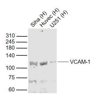 Sample:
Lane 1: Siha (Human) Cell Lysate at 30 ug Lane 2: Huvec (Human) Cell Lysate at 30 ug Lane 3: U251 (Human) Cell Lysate at 30 ug Primary: Anti-VCAM-1 (bs-0396R) at 1/1000 dilution Secondary: IRDye800CW Goat Anti-Rabbit IgG at 1/20000 dilution Predicted band size: 110 kD Observed band size: 110 kD 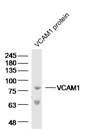 Sample: VCAM1 protein (Human) at 100 ng
Primary: Anti-VCAM-1 (bs-0396R) at 1/300 dilution Secondary: IRDye800CW Goat Anti-Rabbit IgG at 1/20000 dilution Predicted band size: 81 kD Observed band size: 81 kD 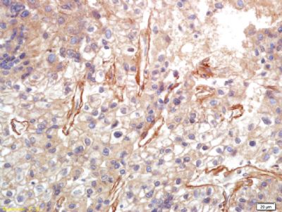 Tissue/cell: human lung carcinoma; 4% Paraformaldehyde-fixed and paraffin-embedded;
Antigen retrieval: citrate buffer ( 0.01M, pH 6.0 ), Boiling bathing for 15min; Block endogenous peroxidase by 3% Hydrogen peroxide for 30min; Blocking buffer (normal goat serum,C-0005) at 37℃ for 20 min; Incubation: Anti-VCAM-1 Polyclonal Antibody, Unconjugated(bs-0396R) 1:200, overnight at 4°C, followed by conjugation to the secondary antibody(SP-0023) and DAB(C-0010) staining 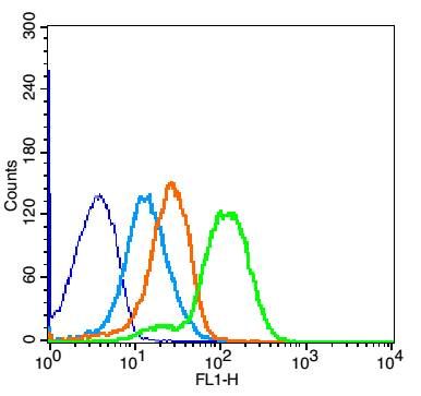 Blank control: Mouse Spleen(blue).
Primary Antibody:Rabbit Anti-VCAM-1 antibody (bs-0396R,Green); Dilution: 1μg in 100 μL 1X PBS containing 0.5% BSA; Isotype Control Antibody: Rabbit IgG(orange) ,used under the same conditions; Secondary Antibody: Goat anti-rabbit IgG-FITC(white blue), Dilution: 1:200 in 1 X PBS containing 0.5% BSA. Protocol The cells were fixed with 2% paraformaldehyde for 10 min at 37℃. Primary antibody (bs-0396R, 1μg /8x10^5 cells) were incubated for 30 min at room temperature, followed by 1 X PBS containing 0.5% BSA + 1 0% goat serum (1 hour) to block non-specific protein-protein interactions. Then the Goat Anti-rabbit IgG/FITC antibody was added into the blocking buffer mentioned above to react with the primary antibody at 1/200 dilution for 40 min at room temperature. Acquisition of 20,000 events was performed. |