胞质紧密粘连蛋白1/闭锁小带蛋白1抗体
产品名称: 胞质紧密粘连蛋白1/闭锁小带蛋白1抗体
英文名称: ZO-1
产品编号: 1329
产品价格: null
产品产地: 上海
品牌商标: 雅吉
更新时间: null
使用范围: WB ELISA IHC-P IHC-F Flow-Cyt IF
上海雅吉生物科技有限公司
- 联系人 :
- 地址 : 上海市闵行区元江路5500号第1幢5658室
- 邮编 :
- 所在区域 : 上海
- 电话 : 158****3937 点击查看
- 传真 : 点击查看
- 邮箱 : yajikit@163.com
| 中文名称 | 胞质紧密粘连蛋白1/闭锁小带蛋白1抗体 |
| 别 名 | ZO1 tight junction protein; Tight junction protein 1; Tight junction protein ZO-1; Tight junction protein ZO1; TJP1; zo-1; Zo1; ZO1_HUMAN; Zona occludens 1; Zona occludens 1 protein; Zona occludens protein 1; Zonula occludens 1 protein; Zonula occludens protein 1. |
| 研究领域 | 细胞生物 免疫学 信号转导 细胞粘附分子 细胞表面分子 |
| 抗体来源 | Rabbit |
| 克隆类型 | Polyclonal |
| 交叉反应 | Human, Pig, (predicted: Mouse, Rat, Chicken, Dog, Cow, Rabbit, Guinea Pig, ) |
| 产品应用 | WB=1:500-2000 ELISA=1:500-1000 IHC-P=1:100-500 IHC-F=1:100-500 Flow-Cyt=1μg/Test IF=1:100-500 (石蜡切片需做抗原修复) not yet tested in other applications. optimal dilutions/concentrations should be determined by the end user. |
| 分 子 量 | 191kDa |
| 细胞定位 | 细胞浆 细胞膜 |
| 性 状 | Liquid |
| 浓 度 | 1mg/ml |
| 免 疫 原 | KLH conjugated synthetic peptide derived from human ZO-1:1551-1702/1733 |
| 亚 型 | IgG |
| 纯化方法 | affinity purified by Protein A |
| 储 存 液 | 0.01M TBS(pH7.4) with 1% BSA, 0.03% Proclin300 and 50% Glycerol. |
| 保存条件 | Shipped at 4℃. Store at -20 °C for one year. Avoid repeated freeze/thaw cycles. |
| PubMed | PubMed |
| 产品介绍 | This gene encodes a protein located on a cytoplasmic membrane surface of intercellular tight junctions. The encoded protein may be involved in signal transduction at cell-cell junctions. Two transcript variants encoding distinct isoforms have been identified for this gene. The N-terminal may be involved in transducing a signal required for tight junction assembly, while the C-terminal may have specific properties of tight junctions. The alpha domain might be involved in stabilizing junctions. Function: The N-terminal may be involved in transducing a signal required for tight junction assembly, while the C-terminal may have specific properties of tight junctions. The alpha domain might be involved in stabilizing junctions. Plays a role in the regulation of cell migration by targeting CDC42BPB to the leading edge of migrating cells. Subunit: Interacts with BVES (via the C-terminus cytoplasmic tail). Interacts with HSPA4 and KIRREL1. Homodimer, and heterodimer with TJP2/ZO-2 and TJP3/ZO-3. Interacts with OCLN, CALM, claudins, CGN/cingulin, CXADR, GJA12, GJD3 and UBN1. Interacts (via ZU5 domain) with CDC42BPB and MYZAP. Interacts (via PDZ domain) with GJA1. Subcellular Location: Cell membrane; Peripheral membrane protein; Cytoplasmic side. Cell junction, tight junction. Cell junction. Cell junction, gap junction. Note=Moves from the cytoplasm to the cell membrane concurrently with cell-cell contact. Detected at the leading edge of migrating and wounded cells. Tissue Specificity: The alpha-containing isoform is found in most epithelial cell junctions. The short isoform is found both in endothelial cells and the highly specialized epithelial junctions of renal glomeruli and Sertoli cells of the seminiferous tubules. Post-translational modifications: Phosphorylated. Dephosphorylated by PTPRJ. Similarity: Belongs to the MAGUK family. Contains 1 guanylate kinase-like domain. Contains 3 PDZ (DHR) domains. Contains 1 SH3 domain. Contains 1 ZU5 domain. SWISS: Q07157 Gene ID: 7082 Database links: Entrez Gene: 7082 Human Entrez Gene: 21872 Mouse Omim: 601009 Human SwissProt: Q07157 Human SwissProt: P39447 Mouse Unigene: 510833 Human Unigene: 4342 Mouse Important Note: This product as supplied is intended for research use only, not for use in human, therapeutic or diagnostic applications. 胞质紧密粘连蛋白1(ZO-1)是多结构域蛋白家族膜结合鸟苷酸激酶的家族成员,在紧密连接蛋白的组成成分中起到对组织分化和器官形成方面起较重要的作用。ZO-1在包括肾、胎盘、血脑屏障等许多组织都有不同的表达,可与紧密连接上的很多跨膜蛋白相互作用。也有学者认为:ZO-1的改变与细胞通透性的增加有关。 |
| 产品图片 | 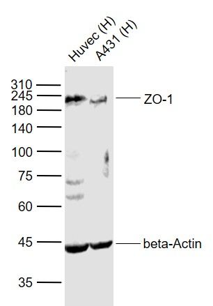 Sample: Sample:Lane 1: Huvec (Human) Cell Lysate at 30 ug Lane 2: A431 (Human) Cell Lysate at 30 ug Primary: Anti-ZO-1 (bs-1329R) at 1/1000 dilution Anti-beta-Actin (bs-0061R) at 1/2000 dilution Secondary: IRDye800CW Goat Anti-Rabbit IgG at 1/20000 dilution Predicted band size: 220 kD Observed band size: 220 kD 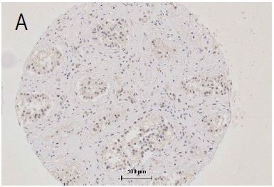 Independently Validated Antibody, image provided by Science Direct, badge number 029577:Formalin-fixed and paraffin embedded human testis labeled with Anti-ZO-1 Polyclonal Antibody, Unconjugated (bs-1329R) at 1:250 followed by conjugation to the secondary antibody. Independently Validated Antibody, image provided by Science Direct, badge number 029577:Formalin-fixed and paraffin embedded human testis labeled with Anti-ZO-1 Polyclonal Antibody, Unconjugated (bs-1329R) at 1:250 followed by conjugation to the secondary antibody.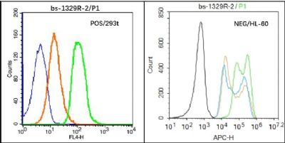 Black line : Positive blank control (293T); Negative blank control (HL60) Black line : Positive blank control (293T); Negative blank control (HL60)Green line : Primary Antibody (Rabbit Anti-ZO-1 antibody (bs-11320R) ) Orange line:Isotype Control Antibody (Rabbit IgG) . Blue line : Secondary Antibody (Goat anti-rabbit IgG-AF647) 293T(Positive)and HL60(Negative control)cells (black) were fixed with 4% PFA for 10min at room temperature, permeabilized with PBST for 20 min at room temperature, and incubated in 5% BSA blocking buffer for 30 min at room temperature. Cells were then stained with ZO-1 Antibody(bs-1329R)at 1:50 dilution in blocking buffer and incubated for 30 min at room temperature, washed twice with 2% BSA in PBS, followed by secondary antibody(blue) incubation for 40 min at room temperature. Acquisitions of 20,000 events were performed. Cells stained with primary antibody (green), and isotype control (orange). 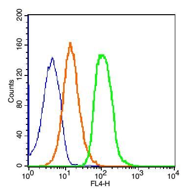 Blank control: 293T Cells(blue). Blank control: 293T Cells(blue).Primary Antibody: Rabbit Anti-ZO-1/AF647 Conjugated antibody (bs-1329R-AF647), Dilution: 1μg in 100 μL 1X PBS containing 0.5% BSA; Isotype Control Antibody: Rabbit IgG/AF647(orange) ,used under the same conditions. Protocol The cells were washed twice with phosphate-buffered saline (PBS). The cells were incubated in 1 X PBS containing 0.5% BSA + 1 0% goat serum (15 min) to block non-specific protein-protein interactions followed by the antibody (bs-1329R-AF647, 1μg /1x10^6 cells) for 30 min on ice. Acquisition of 20,000 events was performed. |