CD13氨肽酶N抗体
产品名称: CD13氨肽酶N抗体
英文名称: CD13
产品编号: 1383
产品价格: null
产品产地: 上海
品牌商标: 雅吉
更新时间: null
使用范围: ELISA IHC-P IHC-F Flow-Cyt IF
上海雅吉生物科技有限公司
- 联系人 :
- 地址 : 上海市闵行区元江路5500号第1幢5658室
- 邮编 :
- 所在区域 : 上海
- 电话 : 158****3937
- 传真 : 021-34661275
- 邮箱 : yajikit@163.com
| 中文名称 | CD13氨肽酶N抗体 |
| 别 名 | ANPEN; Aminopeptidase N; Alanyl aminopeptidase; Alanyl membrane aminopeptidase; Aminopeptidase M; Aminopeptidase N; ANPEP; APN; CD 13; CD13 antigen; gp150; hAPN; Lap 1; Lap1; Alanyl (membrane) aminopeptidase; AMPN_HUMAN; AP M; AP N; AP-M; AP-N; CD13; LAP 1; LAP1; PEPN; Microsomal aminopeptidase; Myeloid plasma membrane glycoprotein CD13; p150 |
| 研究领域 | 肿瘤 细胞生物 干细胞 细胞表面分子 |
| 抗体来源 | Rabbit |
| 克隆类型 | Polyclonal |
| 交叉反应 | Human, Mouse, (predicted: Rat, ) |
| 产品应用 | ELISA=1:500-1000 IHC-P=1:100-500 IHC-F=1:100-500 Flow-Cyt=1µg/Test IF=1:100-500 (石蜡切片需做抗原修复) not yet tested in other applications. optimal dilutions/concentrations should be determined by the end user. |
| 分 子 量 | 109kDa |
| 细胞定位 | 细胞膜 |
| 性 状 | Liquid |
| 浓 度 | 1mg/ml |
| 免 疫 原 | KLH conjugated synthetic peptide derived from human CD13:344-444/444 |
| 亚 型 | IgG |
| 纯化方法 | affinity purified by Protein A |
| 储 存 液 | 0.01M TBS(pH7.4) with 1% BSA, 0.03% Proclin300 and 50% Glycerol. |
| 保存条件 | Shipped at 4℃. Store at -20 °C for one year. Avoid repeated freeze/thaw cycles. |
| PubMed | PubMed |
| 产品介绍 | Broad specificity aminopeptidase. Plays a role in the final digestion of peptides generated from hydrolysis of proteins by gastric and pancreatic proteases. May play a critical role in the pathogenesis of cholesterol gallstone disease. May be involved in the metabolism of regulatory peptides of diverse cell types including small intestinal and tubular epithelial cells, macrophages, granulocytes and synaptic membranes from the CNS. Found to cleave antigen peptides bound to major histocompatibility complex class II molecules of presenting cells and to degrade neurotransmitters at synaptic junctions. Is also implicated as a regulator of IL-8 bioavailability in the endometrium, and therefore may contribute to the regulation of angiogenesis. Is used as a marker for acute myeloid leukemia and plays a role in tumor invasion. In case of human coronavirus 229E (HCoV-229E) infection, serves as receptor for HCoV-229E spike glycoprotein. Mediates as well human cytomegalovirus (HCMV) infection. Expressed in epithelial cells. Belongs to the peptidase M1 family. Function: Broad specificity aminopeptidase. Plays a role in the final digestion of peptides generated from hydrolysis of proteins by gastric and pancreatic proteases. May play a critical role in the pathogenesis of cholesterol gallstone disease. May be involved in the metabolism of regulatory peptides of diverse cell types, responsible for the processing of peptide hormones, such as angiotensin III and IV, neuropeptides, and chemokines. Found to cleave antigen peptides bound to major histocompatibility complex class II molecules of presenting cells and to degrade neurotransmitters at synaptic junctions. Is also implicated as a regulator of IL-8 bioavailability in the endometrium, and therefore may contribute to the regulation of angiogenesis. Is used as a marker for acute myeloid leukemia and plays a role in tumor invasion. In case of human coronavirus 229E (HCoV-229E) infection, serves as receptor for HCoV-229E spike glycoprotein. Mediates as well human cytomegalovirus (HCMV) infection. Subunit: Homodimer. Interacts with the S1 domain of HCoV-229E spike protein. Subcellular Location: Cell membrane; Single-pass type II membrane protein. Cytoplasm, cytosol (Potential). Note=A soluble form has also been detected. Tissue Specificity: Expressed in epithelial cells of the kidney, intestine, and respiratory tract; granulocytes, monocytes, fibroblasts, endothelial cells, cerebral pericytes at the blood-brain barrier, synaptic membranes of cells in the CNS. Also expressed in endometrial stromal cells, but not in the endometrial glandular cells. Found in the vasculature of tissues that undergo angiogenesis and in malignant gliomas and lymph node metastases from multiple tumor types but not in blood vessels of normal tissues. A soluble form has been found in plasma. It is found to be elevated in plasma and effusions of cancer patients. Post-translational modifications: Sulfated. N- and O-glycosylated. May undergo proteolysis and give rise to a soluble form. Similarity: Belongs to the peptidase M1 family. SWISS: P15144 Gene ID: 290 Database links: Entrez Gene: 290 Human Entrez Gene: 16790 Mouse Entrez Gene: 81641 Rat Omim: 151530 Human SwissProt: P15144 Human SwissProt: P97449 Mouse SwissProt: P15684 Rat Unigene: 1239 Human Unigene: 4487 Mouse Unigene: 11132 Rat Unigene: 179371 Rat Important Note: This product as supplied is intended for research use only, not for use in human, therapeutic or diagnostic applications. 氨肽酶N(又称CD13)是氨肽酶系列的一种,氨肽酶是从蛋白质多肽链氨基端催化降解氨基酸残基的水解蛋白酶。是多种冠状病毒的受体,目前在肿瘤侵袭、转移、免疫调节和病毒感染等多方面受人们的关注.它在肿瘤细胞表面高水平表达,对肿瘤细胞外基底膜起到降解作用引发肿瘤的侵袭和转移。主要分布于小肠和肾脏,巨噬细胞、粒细胞和中枢神经系统的突触膜也表达CD13. |
| 产品图片 | 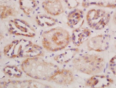 Tissue/cell: mouse kidney tissue; 4% Paraformaldehyde-fixed and paraffin-embedded; Tissue/cell: mouse kidney tissue; 4% Paraformaldehyde-fixed and paraffin-embedded;Antigen retrieval: citrate buffer ( 0.01M, pH 6.0 ), Boiling bathing for 15min; Block endogenous peroxidase by 3% Hydrogen peroxide for 30min; Blocking buffer (normal goat serum,C-0005) at 37℃ for 20 min; Incubation: Anti-CD13/APN/ANPEN Polyclonal Antibody, Unconjugated(bs-1383R) 1:200, overnight at 4°C, followed by conjugation to the secondary antibody(SP-0023) and DAB(C-0010) staining 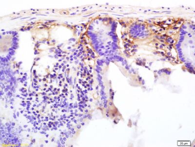 Tissue/cell: mouse colon tissue; 4% Paraformaldehyde-fixed and paraffin-embedded; Tissue/cell: mouse colon tissue; 4% Paraformaldehyde-fixed and paraffin-embedded;Antigen retrieval: citrate buffer ( 0.01M, pH 6.0 ), Boiling bathing for 15min; Block endogenous peroxidase by 3% Hydrogen peroxide for 30min; Blocking buffer (normal goat serum,C-0005) at 37℃ for 20 min; Incubation: Anti-CD13/APN/ANPEN Polyclonal Antibody, Unconjugated(bs-1383R) 1:200, overnight at 4°C, followed by conjugation to the secondary antibody(SP-0023) and DAB(C-0010) staining 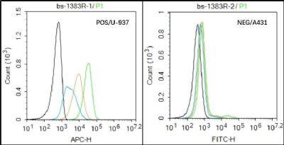 Black line : Positive blank control U937); Negative blank control (A431) Black line : Positive blank control U937); Negative blank control (A431)Green line : Primary Antibody (Rabbit Anti- CD13 antibody (bs-1383R) ) Orange line:Isotype Control Antibody (Rabbit IgG) . Blue line : Secondary Antibody (Goat anti-rabbit IgG-AF647) U937(Positive)and A431 Negative control)cells (black) were incubated in 5% BSA blocking buffer for 30 min at room temperature. Cells were then stained with CD13 Antibody(bs-1383R)at 1:100 dilution in blocking buffer and incubated for 30 min at room temperature, washed twice with 2% BSA in PBS, followed by secondary antibody(blue) incubation for 40 min at room temperature. Acquisitions of 20,000 events were performed. Cells stained with primary antibody (green), and isotype control (orange). 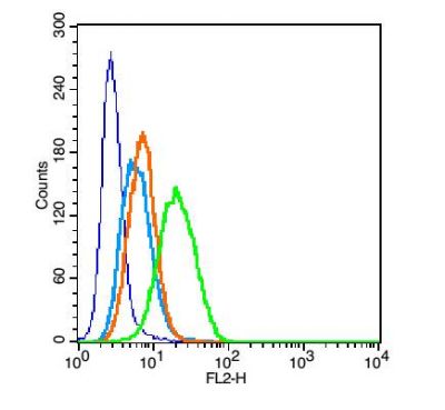 Blank control: U937 (blue). Blank control: U937 (blue).Primary Antibody:Rabbit Anti- CD13 antibody(bs-1383R), Dilution: 1μg in 100 μL 1X PBS containing 0.5% BSA; Isotype Control Antibody: Rabbit IgG(orange) ,used under the same conditions ); Secondary Antibody: Goat anti-rabbit IgG-PE(white blue), Dilution: 1:200 in 1 X PBS containing 0.5% BSA. Protocol The cells were fixed with 2% paraformaldehyde (10 min). Primary antibody (bs-1383R, 1μg /1x10^6 cells) were incubated for 30 min on the ice, followed by 1 X PBS containing 0.5% BSA + 1 0% goat serum (15 min) to block non-specific protein-protein interactions. Then the Goat Anti-rabbit IgG/PE antibody was added into the blocking buffer mentioned above to react with the primary antibody at 1/200 dilution for 30 min on ice. Acquisition of 20,000 events was performed. |