Bcl-xL蛋白抗体
产品名称: Bcl-xL蛋白抗体
英文名称: Bcl-xL
产品编号: 1336
产品价格: null
产品产地: 上海
品牌商标: 雅吉
更新时间: null
使用范围: WB ELISA IHC-P IHC-F Flow-Cyt ICC IF
- 联系人 :
- 地址 : 上海市闵行区元江路5500号第1幢5658室
- 邮编 :
- 所在区域 : 上海
- 电话 : 158****3937 点击查看
- 传真 : 点击查看
- 邮箱 : yajikit@163.com
| 中文名称 | Bcl-xL蛋白抗体 |
| 别 名 | Apoptosis regulator Bcl X; Apoptosis regulator Bcl-X; Apoptosis regulator BclX; B cell lymphoma 2 like; B2CL1_HUMAN; Bcl 2 like 1 protein; Bcl X; Bcl xL; BCL XL/S; Bcl xS; Bcl-2-like protein 1; Bcl2 Like 1; Bcl2 related gene; Bcl2-L-1; BCL2L; Bcl2l1; BCLX; BclXL; BclXs; DKFZp781P2092; PPP1R52; Protein phosphatase 1 regulatory subunit 52; |
研究领域肿瘤 细胞生物 免疫学 信号转导 细胞凋亡 转录调节因子 线粒体
抗体来源Rabbit
克隆类型Polyclonal
交叉反应Human, Mouse, (predicted: Rat, Dog, Pig, Horse, Sheep, )
产品应用WB=1:500-2000 ELISA=1:500-1000 IHC-P=1:100-500 IHC-F=1:100-500 Flow-Cyt=1ug/Test ICC=1:100-500 IF=1:100-500 (石蜡切片需做抗原修复)
not yet tested in other applications.
optimal dilutions/concentrations should be determined by the end user.
分 子 量26kDa
细胞定位细胞核 细胞浆 细胞膜 线粒体
性 状Liquid
浓 度1mg/ml
免 疫 原KLH conjugated synthetic peptide derived from human Bcl-xL:81-200/233
亚 型IgG
纯化方法affinity purified by Protein A
储 存 液0.01M TBS(pH7.4) with 1% BSA, 0.03% Proclin300 and 50% Glycerol.
保存条件Shipped at 4℃. Store at -20 °C for one year. Avoid repeated freeze/thaw cycles.
PubMedPubMed
产品介绍The protein encoded by this gene belongs to the BCL-2 protein family. BCL-2 family members form hetero- or homodimers and act as anti- or pro-apoptotic regulators that are involved in a wide variety of cellular activities. The proteins encoded by this gene are located at the outer mitochondrial membrane, and have been shown to regulate outer mitochondrial membrane channel (VDAC) opening. VDAC regulates mitochondrial membrane potential, and thus controls the production of reactive oxygen species and release of cytochrome C by mitochondria, both of which are the potent inducers of cell apoptosis. Two alternatively spliced transcript variants, which encode distinct isoforms, have been reported. The longer isoform acts as an apoptotic inhibitor and the shorter form acts as an apoptotic activator. [provided by RefSeq, Jul 2008].
Function:
Potent inhibitor of cell death. Inhibits activation of caspases (By similarity). Appears to regulate cell death by blocking the voltage-dependent anion channnel (VDAC) by binding to it and preventing the release of the caspase activator, CYC1, from the mitochondrial membrane. Also acts as a regulator of G2 checkpoint and progression to cytokinesis during mitosis.
Isoform Bcl-X(S) promotes apoptosis.
Subunit:
Homodimer. Isoform Bcl-X(L) forms heterodimers with BAX, BAK or BCL2. Heterodimerization with BAX does not seem to be required for anti-apoptotic activity. Interacts with BCL2L11. Interacts with DMN1L; the interaction stimulates the GTPase activity of DMN1L in synapses and increases the number of axonal mitochondria and the size and number of synaptic vesicle clusters. Interacts with BAD and BBC3. Interacts (isoform Bcl-X(L)) with SIVA1 (isoform 1); the interaction inhibits the anti-apoptotic activity. Interacts with BECN1 and PGAM5. Interacts (isoform Bcl-X(L)) with BAX (isoform Sigma). Isoform Bcl-X(L) interacts with IKZF3. Interacts with HEBP2.
Subcellular Location:
Mitochondrion membrane; Single-pass membrane protein. Nucleus membrane; Single-pass membrane protein; Cytoplasmic side. Cytoplasm, cytoskeleton, centrosome. Note=Mitochondrial membranes and perinuclear envelope. Localizes to the centrosome when phosphorylated at Ser-49.
Tissue Specificity:
Bcl-X(S) is expressed at high levels in cells that undergo a high rate of turnover, such as developing lymphocytes. In contrast, Bcl-X(L) is found in tissues containing long-lived postmitotic cells, such as adult brain.
Post-translational modifications:
Proteolytically cleaved by caspases during apoptosis. The cleaved protein, lacking the BH4 motif, has pro-apoptotic activity.
Phosphorylated on Ser-62 by CDK1. This phosphorylation is partial in normal mitotic cells, but complete in G2-arrested cells upon DNA-damage, thus promoting subsequent apoptosis probably by triggering caspases-mediated proteolysis. Phosphorylated by PLK3, leading to regulate the G2 checkpoint and progression to cytokinesis during mitosis. Phosphorylation at Ser-49 appears during the S phase and G2, disappears rapidly in early mitosis during prometaphase, metaphase and early anaphase, and re-appears during telophase and cytokinesis.
Similarity:
Belongs to the Bcl-2 family.
SWISS:
Q07817
Gene ID:
598
Database links:
Entrez Gene: 598 Human
Entrez Gene: 12048 Mouse
Entrez Gene: 24888 Rat
Omim: 600039 Human
SwissProt: Q07817 Human
SwissProt: Q64373 Mouse
SwissProt: P53563 Rat
Unigene: 516966 Human
Unigene: 238213 Mouse
Unigene: 10323 Rat
Important Note:
This product as supplied is intended for research use only, not for use in human, therapeutic or diagnostic applications.
Bcl X/L蛋白是Bcl蛋白家族的成员之一,是细胞中抑制细胞凋亡的重要分子之一,Bcl-X/L是结构上与Bcl-2具有43%同源性的蛋白,与Bcl-2的作用相同,可抑制细胞凋亡,在肿瘤的发生和发展中起重要作用。
| 产品图片 |
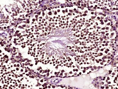 Paraformaldehyde-fixed, paraffin embedded (mouse testis tissue); Antigen retrieval by boiling in sodium citrate buffer (pH6.0) for 15min; Block endogenous peroxidase by 3% hydrogen peroxide for 20 minutes; Blocking buffer (normal goat serum) at 37°C for 30min; Antibody incubation with (Bcl-xL) Polyclonal Antibody, Unconjugated (bs-1336R) at 1:400 overnight at 4°C, followed by operating according to SP Kit(Rabbit) (sp-0023) instructionsand DAB staining.
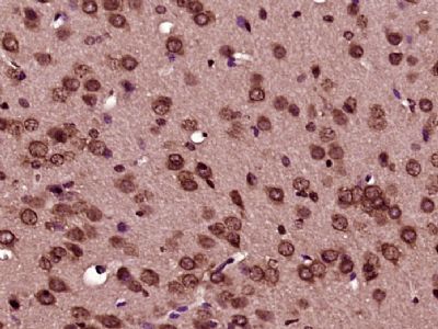 Paraformaldehyde-fixed, paraffin embedded (mouse brain tissue); Antigen retrieval by boiling in sodium citrate buffer (pH6.0) for 15min; Block endogenous peroxidase by 3% hydrogen peroxide for 20 minutes; Blocking buffer (normal goat serum) at 37°C for 30min; Antibody incubation with (Bcl-xL) Polyclonal Antibody, Unconjugated (bs-1336R) at 1:400 overnight at 4°C, followed by operating according to SP Kit(Rabbit) (sp-0023) instructionsand DAB staining.
 Paraformaldehyde-fixed, paraffin embedded (mouse transplanted tumor); Antigen retrieval by boiling in sodium citrate buffer (pH6.0) for 15min; Block endogenous peroxidase by 3% hydrogen peroxide for 20 minutes; Blocking buffer (normal goat serum) at 37°C for 30min; Antibody incubation with (Bcl-xL) Polyclonal Antibody, Unconjugated (bs-1336R) at 1:400 overnight at 4°C, followed by a conjugated secondary (sp-0023) for 20 minutes and DAB staining.
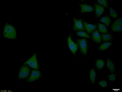 Blank control: A431.
Primary Antibody (green line): Rabbit Anti-Bcl-xL antibody (bs-1336R) Dilution: 1μg /10^6 cells; Isotype Control Antibody (orange line): Rabbit IgG . Secondary Antibody : Goat anti-rabbit IgG-AF647 Dilution: 1μg /test. Protocol The cells were fixed with 4% PFA (10min at room temperature)and then permeabilized with 90% ice-cold methanol for 20 min at-20℃. The cells were then incubated in 5%BSA to block non-specific protein-protein interactions for 30 min at at room temperature .Cells stained with Primary Antibody for 30 min at room temperature. The secondary antibody used for 40 min at room temperature. Acquisition of 20,000 events was performed. |